

When we talk about a membrane being “permeable,” we usually mean two different things and use one word for both. Diffusive permeability is how easily molecules cross the membrane by diffusion. Hydraulic permeability is how easily water flows through it when there is a pressure difference across it. If you are interested in filtration, you want hydraulic permeability as high as possible. For cell culture we want the diffusive permeability to be as high as possible and the hydraulic permeability to be as low as possible. Why? To avoid coupling the fluid in one compartment to the cells in the other.
Two kinds of permeability
Our ultrathin membranes are famous for diffusive permeability. A 100 nm thick membrane offers almost no resistance to small molecules, so a cell on NPN sees its basal medium nearly as if the membrane were not there.
Hydraulic permeability (Lp) has units of flow per area per pressure, which we report as mL min−1 cm−2 bar−1. Multiply it by a pressure difference and you get a flux of water through the membrane. In a culture device that pressure difference is never zero. A 1 mm mismatch between the liquid level in a top well and a bottom channel is about 10 Pa. Pipetting creates heads of centimeters, briefly.
How we predict it
For a pore of radius a in a membrane of thickness L, the resistance to flow has two parts in series: an entrance and exit term (flow through a hole in a thin plate) and a Poiseuille term for the length of the pore. Dagan, Weinbaum and Pfeffer (1982) gave the combined result:
ΔP = (μQ / a3) · [3 + 8L / (πa)]
For our membranes L and a are about the same size, so the entrance term matters as much as the length term. That is why ultrathin membranes beat the predictions of conventional membrane theory, and it is the model behind the permeability predictions in DesOrmeaux et al. (2014). Summing the flow through every pore and dividing by area gives Lp.
What the numbers look like for different membranes we make
| Membrane | Pore size | Porosity | Thickness | Lp (mL min−1 cm−2 bar−1) |
|---|---|---|---|---|
| Track-etched | comparable to NPN | low | microns | ~0.3 |
| NPN | ~60 nm | ~15% | 100 nm | ~75 |
| Dual scale, low porosity | 3 µm on NPN | 0.7% + 15% | 100 nm | ~700 |
| ibidi ibiPore SiN | 0.5 µm | 20% | 400 nm | ~1,350 |
| ibidi ibiPore SiN | 3 µm | 5% | 400 nm | ~3,900 |
| ibidi ibiPore SiN | 8 µm | 5% | 400 nm | ~11,700 |
These are ideal membrane-only predictions (water, 1 mPa·s); a real device adds resistance of its own. The track-etched value is a vendor figure quoted in DesOrmeaux et al.
Two things stand out. Micropores dominate: in the low porosity dual scale membrane, 0.7% of the area carries about 90% of the flow, because the flow through an open hole scales with the cube of its radius. And everything with micron-scale pores sits well above physiological interstitial flow (about 0.1 to 1 µm/s) at a head of only a millimeter.
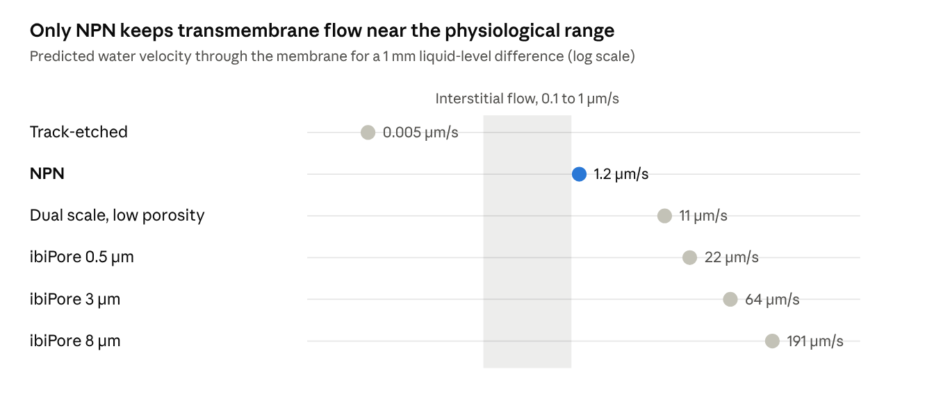
At the same 1 mm head, micron-scale pores push roughly 10 to 150 times more fluid through the cell layer than NPN does.
The good side: filtration (Gaborski 2010, Winans 2016)
For a filter, high hydraulic permeability is the whole point. Tom Gaborski’s 2010 ACS Nano paper measured water flow through 15 nm pnc-Si and found it matched the Dagan model across five wafers from 0.7% to 13.5% porosity. At 15% porosity the membranes passed about 40 mL/(cm²·min·bar), the highest then reported for a membrane with pores under 100 nm. That is about 20 times a commercial ultrafiltration membrane and more than 35 times a 6 µm track-etched membrane with similar pores.
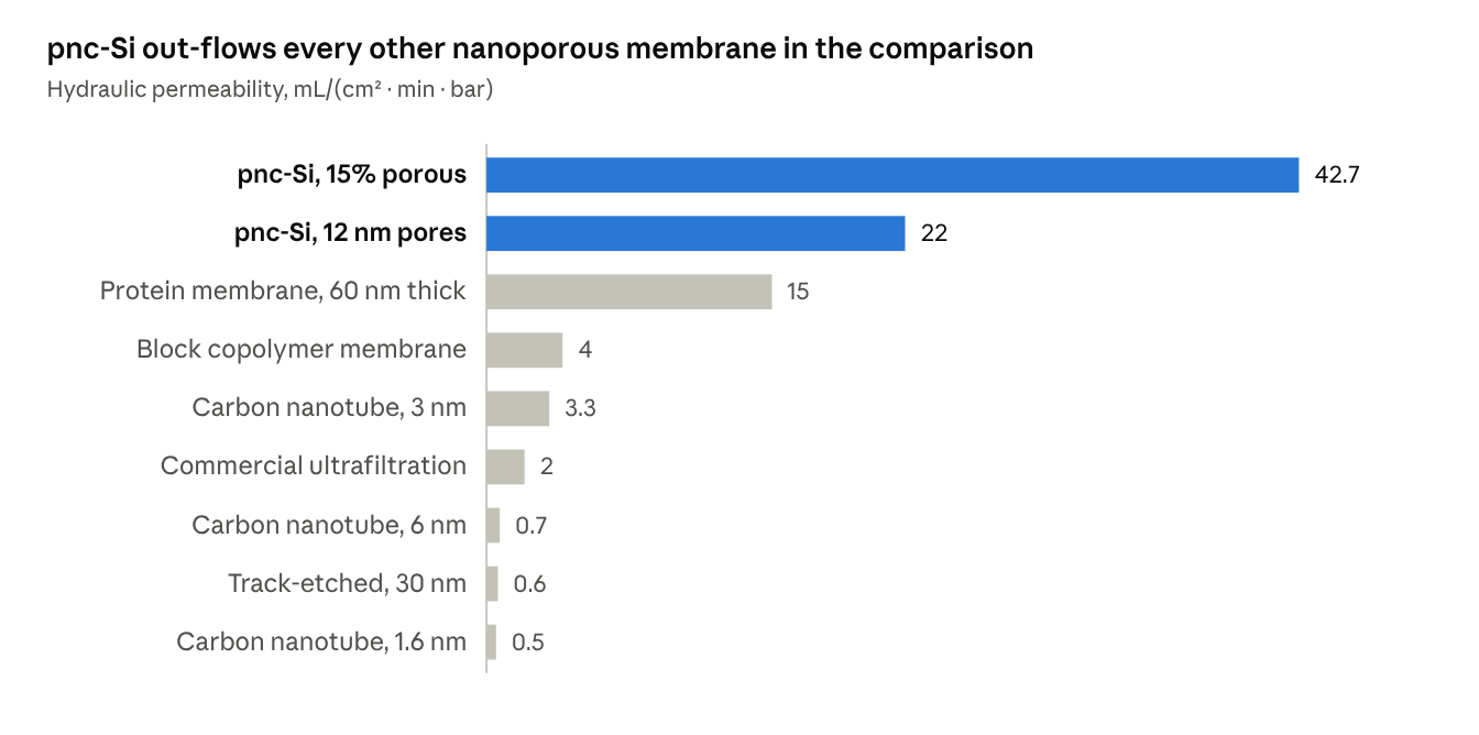
Winans et al. (2016) showed the cost. In dead-end filtration of 20 nm particles, NPN started more than two orders of magnitude more permeable than track-etched membranes, then fouled quickly: the fast flux packed rejected particles into a cake on the membrane. Slowing the centrifuge from 2000 to 500 rpm raised NPN’s capacity 8-fold, to match track-etched, while its permeability stayed orders of magnitude higher. The advantage is speed: practical volumes processed in a short time at low pressure.
The property that makes a filter fast is the same one that pushes medium through a cell layer. Which one you get depends on whether you want flow through the membrane.
Why it matters in culture
Once a tight monolayer forms, the cells themselves are the dominant resistance and the membrane drops out of the problem. The membrane matters before that, and wherever the monolayer is incomplete:
- Seeding. Medium draining through the membrane pulls cells onto pores, and through them when the pores are microns wide. On the more open membranes that drainage is as fast as cells settle.
- Medium changes. Every pipetting step sends a pulse of flow through the cell layer. If the hydraulic permeability is too high, a medium exchange from below can lift cells off the membrane, and a pressurized flow above can crush cells against it.
- Defects. At a gap in the monolayer or an exposed micropore, the whole pressure drop concentrates into a jet.
- Permeability assays. Any level mismatch adds convection to a small molecule permeability measurement, so a “leaky” barrier can partly be a pressure artifact.
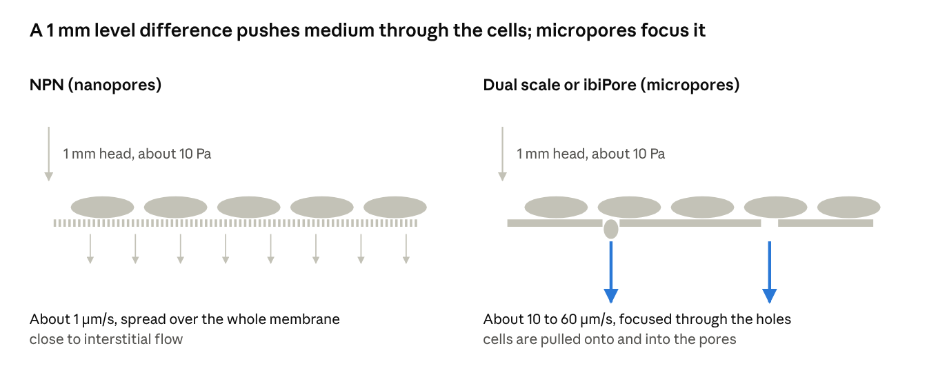
Before a monolayer closes, the same small head drives about ten times more flow through a micropored membrane, and that flow concentrates at the holes.
The bad side: crushing cells with flow (Salminen et al., 2019)
Alec Salminen built dual-scale membranes with a gradient of eight 3 µm pore spacings (6 to 27 µm center to center) on one nanoporous membrane, so every density sat in a single field of view (Figure 5 of the paper). After 24 h of shear at 4.5 dyn/cm², endothelial cells were lost from the densest region: 787 cells/mm² at 27,777 pores/mm², against 1,172 cells/mm² at 1,371 pores/mm² (Figure 5C). That is about 56 micropores under each cell at the dense end and about 3 at the sparse end.
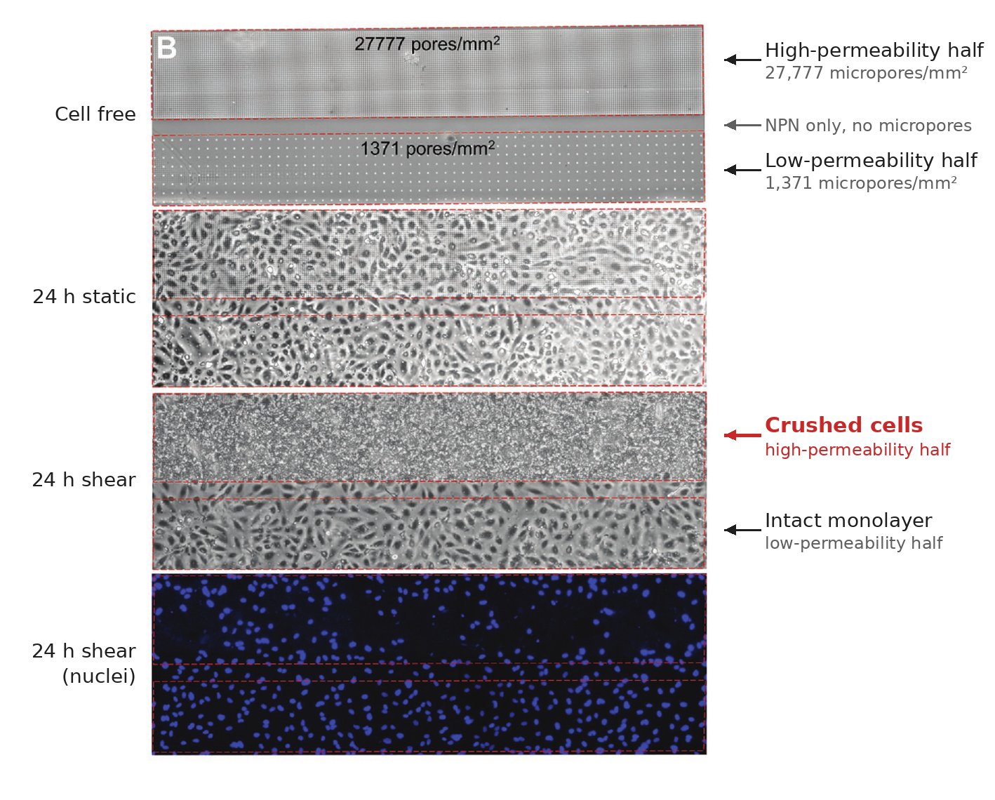
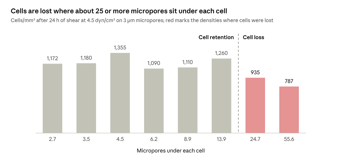
A collagen I gel filling the micropores from below restored adhesion at every density (Figure 6). Because both halves sit on the same membrane under the same flow, the comparison isolates permeability: under pressurized flow above, cells on the high-permeability half are crushed against the membrane, and the monolayer on the low-permeability half stays intact.
The best illustration of hydraulic decoupling: shear-free chemotaxis (Chung et al., 2014)
The clearest demonstration of what a low hydraulic permeability buys you is our 2014 Lab on a Chip paper. Henry Chung built a two-chamber device: a flow chamber that carried a chemoattractant gradient, separated by a 15 nm pnc-Si membrane from a cell chamber where neutrophils crawled. The goal was to deliver the gradient to the cells without exposing them to the shear of the flow.
The analysis treated the membrane as a ladder of pores, each a resistor connecting the two chambers. The result that matters here is in its Figure 3a: nanopores couple the two chambers far less than micropores at the same porosity, because each nanopore is a large hydraulic resistance. Nanoparticle tracking in the cell chamber showed purely diffusive motion with up to 50 µL/min in the flow chamber, and the analysis predicted roughly a 240,000-fold reduction in wall shear across the membrane. Meanwhile the membrane passed small molecules freely, so neutrophils migrated up the fMLP gradient with no bias in the flow direction (about 80% of roughly 400 tracked cells), and cells could be labeled through the membrane in minutes.
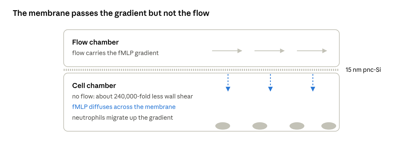
Flow stays in the upper chamber; only molecules reach the neutrophils below.
The test is the paper’s Figure 5. Nanoparticles (210 nm) tracked in the cell chamber moved by Brownian motion alone at flow-chamber rates below 50 µL/min. At 50 µL/min a drift of about 13 µm/min appeared, about what diffusion covers over the 3 min of tracking.
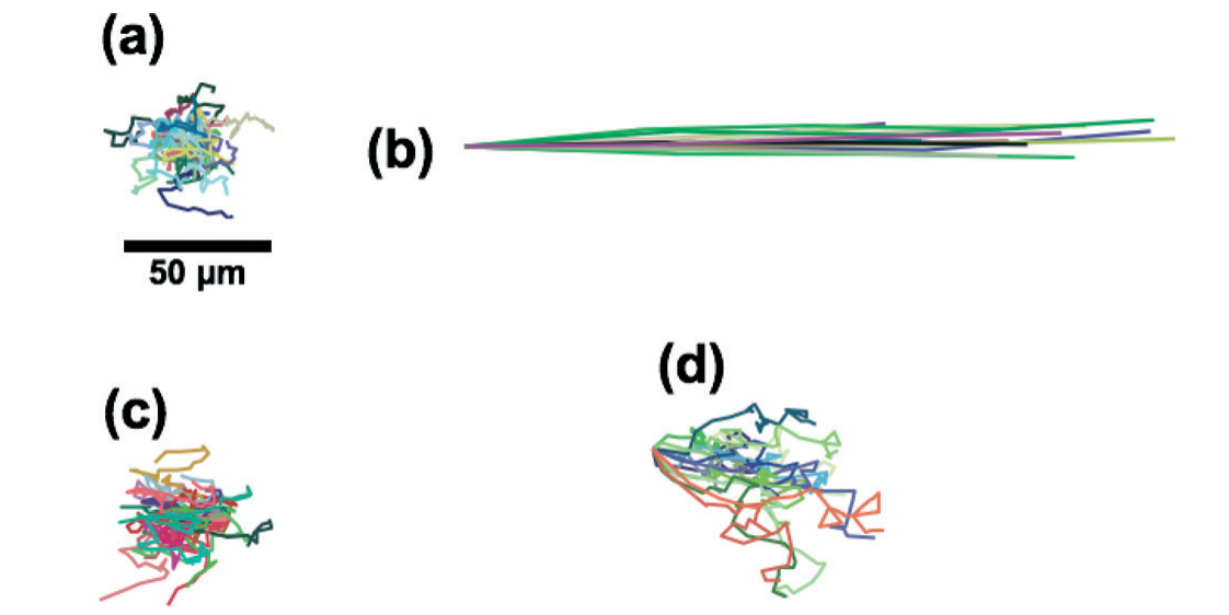
That is the combination we want for culture membranes generally: very high diffusive permeability with a hydraulic permeability low enough that pressure differences do not push fluid through the cells.
Design rule
Choose a membrane whose hydraulic permeability keeps the transmembrane velocity near 1 µm/s for the pressure heads your handling actually creates. NPN already sits there. Adding micropores raises hydraulic permeability by an order of magnitude or more, which is worth knowing whenever we compare results across membranes, and worth controlling for when micropores are the variable of interest.
References
- Gaborski TR, Snyder JL, Striemer CC, Fang DZ, Hoffman M, Fauchet PM, McGrath JL. High performance separation of nanoparticles using ultrathin pnc-Si membranes. ACS Nano 2010;4:6973–6981.
- DesOrmeaux J-PS, Winans JD, Wayson SE, Gaborski TR, Khire TS, Striemer CC, McGrath JL. Nanoporous silicon nitride membranes fabricated from porous nanocrystalline silicon templates. Nanoscale 2014;6:10798–10805.
- Chung HH, Chan CK, Khire TS, Marsh GA, Clark A Jr, Waugh RE, McGrath JL. Highly permeable silicon membranes for shear free chemotaxis and rapid cell labeling. Lab Chip 2014;14:2456–2468.
- Winans JD, Smith KJP, Gaborski TR, Roussie JA, McGrath JL. Membrane capacity and fouling mechanisms for ultrathin nanomembranes in dead-end filtration. J Membr Sci 2016;499:282–289.
- Salminen AT, Zhang J, Madejski GR, Khire TS, Waugh RE, McGrath JL, Gaborski TR. Ultrathin dual-scale nano- and microporous membranes for vascular transmigration models. Small 2019;15:1804111. doi:10.1002/smll.201804111
- Dagan Z, Weinbaum S, Pfeffer R. An infinite-series solution for the creeping motion through an orifice of finite length. J Fluid Mech 1982;115:505–523.
- ibidi µ-Slide ibiPore SiN instructions (IN_852X6): pore sizes, porosity and 400 nm thickness.

Professor McGrath holds a BS degree in Mechanical Engineering from Arizona State and a MS degree in Mechanical Engineering from MIT. He earned a PhD in Biological Engineering from Harvard/MIT's Division of Health Sciences and Technology. He then trained as a Distinguished Post-doctoral Fellow in the Department of Biomedical Engineering at the Johns Hopkins University. Professor McGrath has been on the Biomedical Engineering faculty at the University of Rochester since 2001 where he also served as the director of the graduate program in BME for more than a decade and currently serves as Associate Director of the URNano microfab and metrology core. Professor McGrath also has faculty affiliations with many other programs at UR including the Material Research Program, the Environmental Health and Sciences Center, the Biochemistry and Biophysics program, and the Musculoskeletal Research Center. McGrath's graduate, post-doctoral, and early faculty research was focused on quantitative experiments and mathematical modeling of cell migration covering molecular, cellular, and multi-cellular phenomena. This was true until 2007 when he, along with Professor Philippe Fauchet (now Dean at Vanderbilt) and PhD students Tom Gaborski (RIT) and Chris Streimer (Adarza), discovered a means to self-assembled nanopores in 15 nm thick free-standing silicon and demonstrated the remarkable transport properties of the new material in a Nature paper. This seminal discovery led to the creation of the multidisciplinary Nanomembrane Research Group (NRG) and the founding of SiMPore Inc. in the same year. The NRG and SiMPore have been dedicated to the advancement of ultrathin membrane technologies and exploring all of their potential applications ever since. This blog also dates back to 2007 and has had contributions from more than 100 students, faculty, scientists, engineers, and entrepreneurs. It contains over 2,500 pages and posts logging progress large and small over all these years. Yet somehow it feels like we are just getting started.

Leave a Reply