
Modular µSiM Version 2 (V2) Holds ~30 µl of media in the bottom channel. Designed for cocultures, ELISAs, and other assays that are easier with greater channel media volumes. For more surface area and volume information on mod-µSiM V2, see…

Category: μSiM
All things cell culture on the μSiM
Flow demo (T cell interaction with human in vitro BBB model under flow)

20210122.flow-movie-demo I attached a link to a short presentation regarding the flow movie setups. The figures and videos will be explained in detail during the meeting on Friday.
Posted in memBrains
Pore Analysis for Wafer 1383 (Low Porosity Dual Scale)

From SiMPore’s Basecamp thread … These are the nanopore characteristics. So between 14% and 20% porosity across the wafer and between 43 nm and 50 nm average pore size. So a little under-sized. I hope T-Cells and Monocytes can get…
QC on Dual Scale Membrane (lot 1383)_from Bern

We recently received 100 Dual Scale Membrane (lot 1383). As noted in Dan’s post we also had same issues regarding mix up in the labeling. In the gel box, 2 boxes (25 membranes/box) are labeled 0.625% porosity and 2boxes are…
Posted in memBrains
QC on Dual Scale Membranes (Lot 1383)

Recently we have received dual scale (nanoporous and microporous) membranes from SiMPore at two porosities: 0.625% and 1.25%. These feature 3 µm circular pores (Lot 1383). We routinely perform QC checks on membrane deliveries, so the following is report on…
Posted in memBrains
Collagen IV is deposited below human umbilical vein endothelial cells

Introduction My previous post focused on attempts to reproducibly generate collagen IV with human umbilical vein endothelial cells (HUVECs) so that we would be able to establish protocols for basement membrane protein detection and degradation measurement. Following my presentation on…
Simulation and PIV results of the flow module

1. Introduction Fluid flow is an influential component of vascular models for conditioning cells and introducing other components such as leukocytes. In the previous post, we described our modular design for integrating a flow module into the uSiM device. Here,…
Posted in memBrains
Measuring changes in endothelial basement membrane integrity
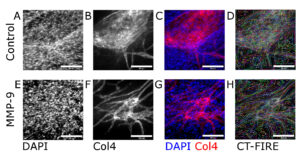
Introduction Inflammatory diseases have the potential to disrupt blood vessel permeability. This has serious ramifications for the blood-brain barrier (BBB), which is the interface between the vasculature and the brain. A healthy BBB allows essential nutrients to pass through while…
Label Free State Analysis and Tracking: Neutrophils on HUVEC Monolayers
Introduction/Methods As part of our investigation into neutrophil transmigration on tissue chip systems featuring HUVEC monolayers, I have been working on developing code for both cell state analysis and tracking using Wolfram Mathematica (and occasionally Python). This project relies on…
Semantic Segmentation for Neutrophils on HUVEC Monolayers: A Machine Learning Approach
Introduction Phase-contrast microscopy is widely utilised in live cell imaging, with pixel segmentation protocols in particular finding widespread usage as a means to computationally isolate cells from a tissue-culture background. Computer segmentation allows for the analysis of individual cell properties…
