
One of the validations of the in vitro human Blood Brain Barrier system is to ensure that the endothelial cells derived from iPSC’s and cultured in our system exhibit behaviors that are consistent with endothelial cell behavior in situ. The…

Category: memBrains
Daily In-Situ Permeability using mouse CMECs

Introduction In the Glading Lab we study Cerebral Cavernous Malformations (CCMs). In CCMs, brain capillaries develop enlarged lumens and a disrupted blood-brain barrier. The presence of a mutation in KRIT1 causes loss of KRIT1 protein function in the endothelial cells.…
iPSC-derived Brain Pericyte-like Cells

Introduction Pericytes are mural cells that line the outer surface of microvessels and regulate diverse aspects of vascular development and function. We differentiate brain-pericyte-like cells (BPLCs) from hiPSC clones using the protocol published by Eric Shusta’s lab (Gastfriend et al.,…
Posted in memBrains
Culture divider insert assembly and test

Introduction We are designing a cell culture chamber insert to allow side-by-side co-culture of cells and ultimately triculture in the µSiM devices. Materials and Methods ALine devices assembly according to the “ALine Modular Device Assembly” instructions. 2-slot NPN membranes applied…
Little Back Dots in Coated Controls … How We Wonder What You Are.

Introduction We are investigating some issues seen with long term attachment (up to 6 days) of the EECM-BMEC-like cells seen by collaborators in Bern, where they noticed black aggregates on the coated control devices that get worse at day 6.…
Using Histogram Distance Measurements as a Means of Analyzing Flow Cytometry Data

Overview Recently, we performed a series of flow cytometry studies assessing neutrophil (PMN) activation in first generation flow devices. All the relevant data and can be found in a previous post: here. As part of the analysis, we chose to…
Utilizing the μSIM to model brain endothelial barrier changes in Cerebral Amyloid Angiopathy

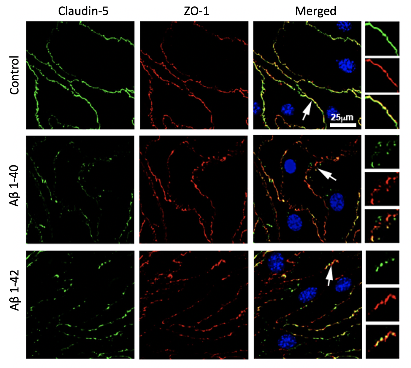
Introduction Vascular hyperpermeability and hemorrhagic stroke are hallmark symptoms of Cerebral Amyloid Angiopathy (CAA), a cerebrovascular disease characterized by amyloid-β deposition around the brain microvasculature. In CAA pathogenesis, vascular Aβ accumulation contributes to structural alterations and loss of integrity in…
Posted in memBrains
AFM Measurements of Glycocalyx in the Upside Down µSiM
Slide deck from Large Group Meeting 12-21-12 (PPT) Access requirements for the AFM cantilver to contact the cells requires the cells to be grown on the membrane surface in what is typically the bottom channel of A-line devices (Typical A-line…
Progress Toward Modeling Inflammation-Mediated Degradation of Basement Membrane Proteins in the μSiM-hNVU

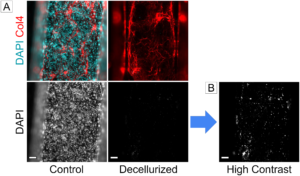
Introduction The blood-brain barrier (BBB) plays an important role in maintaining brain chemistry by restricting the permeability of small molecules and cells. Inflammatory diseases may disrupt the BBB and promote the development of neurological disorders [1]. One potential mechanism of…
Pump-assisted oscillation mixing in μSIM for consistent small volume sampling and measurement from bottom channel.

Background From previous experimental results as well as COMSOL simulations presented in our older posts, we observed a universal non-uniform analyte distribution in the μSIM bottom channel due to limited lateral diffusion rate. This uneven distribution profile will limit our…
Posted in memBrains
